
Unveiling the Art Exhibit of Our Cells
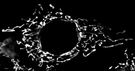
Image of mitochondria stained with MitoRed in a Mouse embryonic fibroblast cell displaying a healthy network. Photo taken by Sophie Hurwitz
When you think of art, what comes to mind? The Mona Lisa, maybe Waterlilies by Monet. Do you ever think about your own cells? Each of our cells is packed full of tiny machines called proteins where the twists and turns of the folds tells us how we each work. The way we look, at literally an atomic level, is a work of art that many of us never get to see.
Since the 1990s, the scientist David Goodsell has been challenging this separation between art and science with watercolors that capture the art of viruses (1 ). These brightly colorful watercolors depict just how packed cells are, as each canvas is almost filled with pastel red and blue paint that represent tons of proteins in viruses and cells. These paintings are based off images of cells that have been obtained through several new methods to picture cells better. These stunning watercolors are a great way to understand how beautiful and complex the small world in each of our cells are.
I got to see this gorgeous world when I imaged the mitochondria inside our cells for the first time. You probably think that when I say mitochondria, I am talking about the small bean shaped powerhouse of the cell. With that image, it’s hard to understand why I would think of the mitochondria as art. However, mitochondria aren’t one or two beans existing in a cell, but a beautiful network that expands from the center of the cell all the way to the edge (2).
Each image of this network unveils an additional work of art, the equilibrium of the processes of fusion and division. ‘Fusion’ refers to how mitochondria can combine to form a network, ‘division’ to how they separate. The balance of these processes helps to maintain beautiful networks in cells and can quickly shift in response to stress. For example, if cells are deprived of sugars, the connected mitochondria will work together and make more energy, leading to a more fully-connected network.
To better understand how these networks are upheld, biochemists study how small proteins regulate fusion and division. Specifically, I study the fusion proteins, which grab onto a new mitochondrion and then pull them together to combine. These proteins are called ‘mitofusins’ (3). To do this, I modify, or ‘mutate’, sections of the mitofusins, causing them to break. I then take pictures of the mitochondria with a fancy microscope to see how the broken mitofusins change the mitochondrial shape. For example, one mutation stops the proteins on one mitochondrion from grabbing and combining to other mitochondria (4). When you image mitochondria with these broken proteins, the network is all totally disconnected!
These photographs are an incredible tool and are a quick way to see the effects of a mutated protein. We can quickly find out which sections of a protein are needed for it to work and potentially even how it works. With increasing methods to image cells and proteins, there will only be a bigger boom in all we can see happening in cells.
Cell images are scientific data, but they’re also the beautiful art of how we all live. This art literally contains knowledge: The more images we get, the more we will be able to reveal about how we all work.
References
(1) David Goodsell: The master of mol art. https://www.asbmb.org/asbmb-today/people/080111/david-goodsell-the-master-of-mol-art (accessed 2024-11-23).
(2) Tilokani, L.; Nagashima, S.; Paupe, V.; Prudent, J. Mitochondrial Dynamics: Overview of Molecular Mechanisms. Essays Biochem. 2018, 62 (3), 341–360. https://doi.org/10.1042/EBC20170104.
(3) Schrepfer, E.; Scorrano, L. Mitofusins, from Mitochondria to Metabolism. Mol. Cell 2016, 61 (5), 683–694. https://doi.org/10.1016/j.molcel.2016.02.022.
(4) Cao, Y.-L.; Meng, S.; Chen, Y.; Feng, J.-X.; Gu, D.-D.; Yu, B.; Li, Y.-J.; Yang, J.-Y.; Liao, S.; Chan, D. C.; Gao, S. Mfn1 Structures Reveal Nucleotide-Triggered Dimerization Critical for Mitochondrial Fusion. Nature 2017, 542 (7641), 372–376. https://doi.org/10.1038/nature21077.