
Diagnosing Liver Cancer in a Snap – Literally!
Have you ever thought to yourself: “Man, I really need to filter my blood after that big meal I just had!” Hopefully the answer to that question is no – if it's no, you have your liver to thank for taking care of that! As our largest (and most awesome) internal organ, the liver is responsible for so many vital processes ranging from digestion to detoxification. However, this means that if anything harmful is to happen to our liver - such as cancer - it can have devastating effects on the rest of the body.
One particular cancer that is challenging and devastating to our liver is called hepatocellular carcinoma, or HCC for short. HCC is the 4th leading cause of cancer-related deaths, partly due to our inability to detect it before significant damage is done. This is because this villainous cancer is difficult to distinguish from other tumors, which are treated for with very different procedures than HCC. Therefore, accurate diagnoses are needed for HCC tumors, and our best evidence for catching a criminal would be photo evidence of them caught in the act. But even with a good image, how can we tell them apart from other liver tumors that are similar shapes and sizes? This is actually done by using the thing the liver deals with the most, blood!
This might prompt even more questions, like how do you take a picture of blood? Thankfully, there is a way to do this with ultrasound – yes the same ones that are used to take pictures of babies in the womb. However, we don't usually “see” blood in normal ultrasound images, it just looks like the black background. With microbubbles, or small little bubbles the size of your red blood cells, we can “see” the blood flow with ultrasound imaging, much like being able to “see” the air currents when blowing bubbles in the backyard. Since 2016, hospitals have actually been using ultrasound with these microbubbles to take pictures of blood flow to these HCC tumors, and identify them. Based on these images, diagnosis is made using a 1-5 scale known as LI-RADS (Liver Reporting and Data System), with a 1 indicating that it is definitely not HCC, and 5 meaning that is definitely is.
This ranking system for liver tumor images might sound like it should be enough to solve this problem and identify HCCs, but it is currently only done by simply observing the images, requiring a lot of background knowledge and experience. It also involves comparing the brightness in between images, which can be very different depending on the settings used to take the picture with ultrasound.
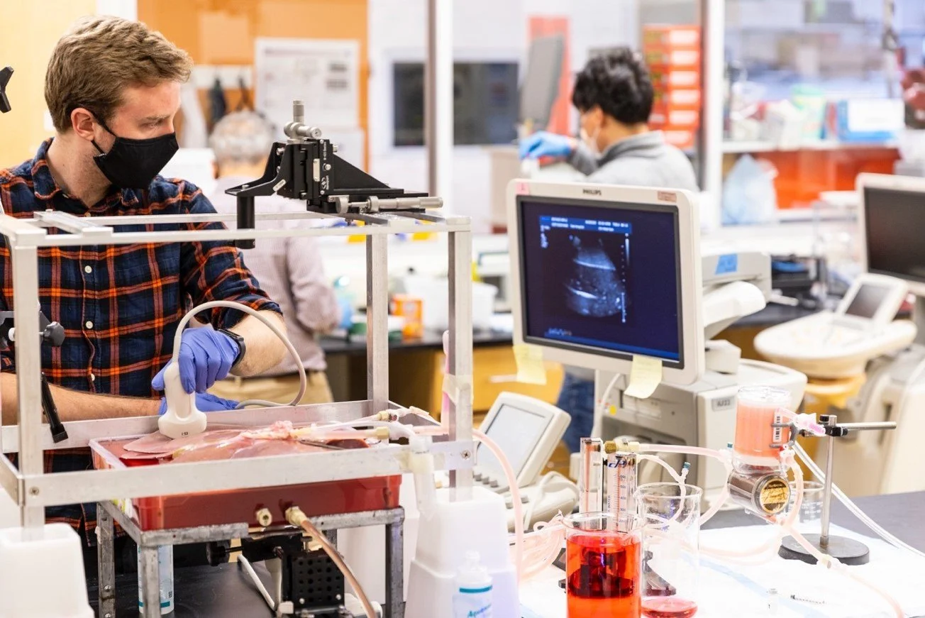
Therefore, here at UW we are developing a new technique to take better pictures of the liver and load these images into a computer script to actually measure the blood flow to the tumor. Just like how a tri-pod can help you get a better picture since you don’t have to worry about your hand shaking, we use a holder called an articulated arm to hold the ultrasound probe, rather than having the doctor hold it. By then giving these improved images to a computer script, it will help to increase the accuracy of diagnosis, since it could provide numbers to doctors that are easier to draw conclusions from than features of the pictures.
Having better pictures and an easier way to analyze them would let us catch these dangerous HCCs faster, and lead to better outcomes for those patients. Plus, with the incredible amount of work our liver does for us, don’t you think it deserves some good pictures?

Connor Krolak is a 2nd year Bioengineering PhD student studying how ultrasound can be used to create better diagnoses for cancer, and then also use ultrasound to treat the cancer as well. He is currently focusing on using ultrasound to diagnose a really difficult type of liver cancer – hepatocellular carcinoma.